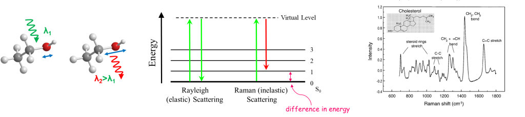
What is Raman Spectroscopy?
Raman is a form of, so-called, vibrational spectroscopy. Raman scattering is observed when a very small number of photons incident on a molecule (about 1 in 10^7) are inelastically scattered and loose or gain a small portion of their energy. A Raman spectrum is a plot of the intensity of scattered light versus the energy difference between incident photons and Raman scattered photons. This energy difference is a result of the energy absorbed or gained from vibrational energy levels which are characteristic of the chemical bonds in the molecules. The Raman spectrum is hence representative of the chemical bonds of the molecules and contains information about the chemical composition and the molecular structure of a substance.
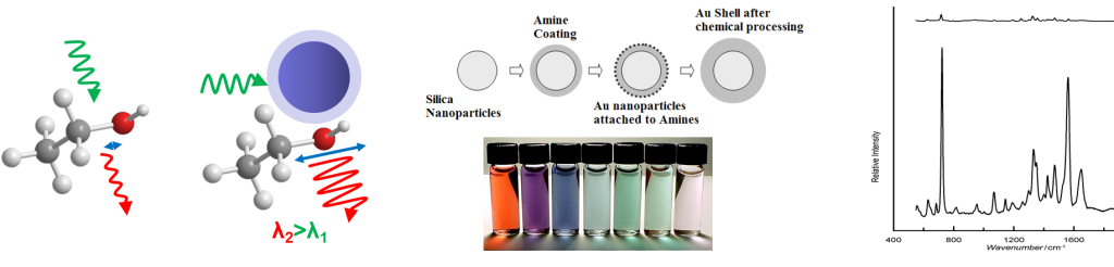
What is Surface Enhanced Raman Spectroscopy SERS?
Surface enhanced Raman Spectroscopy (SERS) is a variation of the technique which offers great enhancement of the, usually extremely weak, Raman signal (up to 10^14). The enhancement is a result of plasmon resonances, the unison oscillation of electrons on the surface of a metallic nanostructure as a result of incident light of the right, resonant frequency. Molecules adjacent to these structures are exposed to strong fields set by these oscillations and produce a strong Raman signal. SERS has had a variety of bioanalytical applications in recent years including the detection and identification of bacteria.
Can we “see and treat” urinary tract infections?
Raman Research at the ODx lab
Urinary tract infections (UTIs) are very common, especially in women, resulting in billions of dollars in health care costs every year. The current, standard, method of diagnosis for a UTI is by quantitative urine culture, which requires 24 hours to produce results. A finding of 10^5 or more colony forming units per ml of urine (cfu/ml) is defined as significant bacteriuria. Antibiotic susceptibility testing requires an additional 24 hour period. Due to the prolonged period of diagnosis, physicians, suspecting an infection, usually prescribe broad spectrum antibiotics before any definitive diagnosis and antibiogram results are obtained. This practice leads to many undesirable consequences such as unsuccessful treatments, chronic infections, rising health care costs, and, most importantly, increased antibiotic resistance. A rapid, accurate, automated and relatively inexpensive method of UTI diagnosis and antibiogram is urgently needed. Development of such a method would result in more specific and more effective treatments and could contribute in reducing microbial antibiotic resistance in the long term. The aim of the work at the ODx Lab is the development of a rapid diagnosis and antibiogram method using SERS spectra obtained from bacteria incubated with various antibiotics and colloidal silver nanoparticles. A successful system should be able to
- Identify the presence of bacteria (quantification)
- Classify the causative bacteria (classification)
- Determine the sensitivity of the bacteria to various antibiotics (classification)
Important characteristics of the proposed system are minimal sample preparation, low cost, and speed.
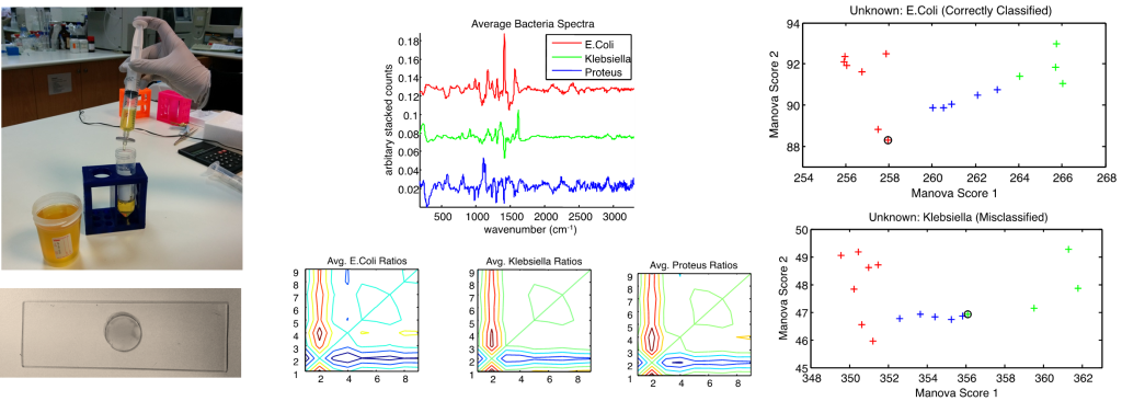
Classification of Bacterial Species Using SERS
Πρόσφατα, η ομάδα μας και άλλοι έχουν δείξει ότι με χρήση φασματοσκοπία Raman και SERS μπορούν να αναγνωρισθούν τα είδη των βακτηρίων. Χρησιμοποιώντας SERS με νανοσωματίδια κολλοειδούς αργύρου, η κατηγοριοποίηση των βακτηριακών ειδών έδωσε ~ 94% ορθό ποσοστό ταξινόμησης (δηλ. μόνο 1 από τα 16 δείγματα ταξινομήθηκε εσφαλμένα).
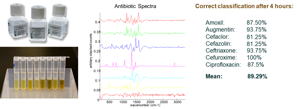
Ευαισθησίa στα Αντιβιοτικά
Ευαισθησία ή αντοχή στα αντιβιοτικά αμοξικιλλίνη, αμοξικιλλίνη / κλαβουλανικό οξύ, κεφαλόρ, κεφαζολίνη, κεφτριαξόνη, κεφουροξίμη και σιπροφλοξασίνη που παρουσιάζονται από Ε. Coli, Κ. Pneumoniae και Proteus spp. προσδιορίστηκαν με ακρίβεια 81-100% χρησιμοποιώντας τα φάσματα SERS μετά από μόλις 2-4 ώρες έκθεσης.
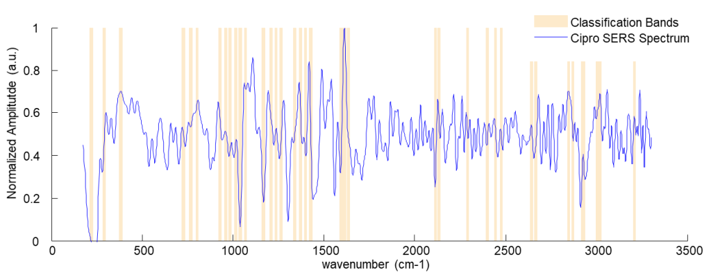
Ευαισθησία στην Ανθεκτικότηα στις Φθοροκινολόνες
In addition, this method was able to distinguish the SERS spectra of bacteria that are treated and are sensitive to ciprofloxacin from bacteria that are resistant, after just a 2 hour incubation time with a correct classification rate of ~ 94%. Ciprofloxacin, unlike other antibiotics previously examined, does not interfere with cell wall synthesis but with DNA synthesis. It appears, from our preliminary results, that the classification of bacteria as sensitive or resistant may have been based on the presence of multi-drug efflux pumps on the cell wall of resistant bacteria which function to remove ciprofloxacin and other newer fluoroquinolones and are suspected to be the major cause of resistance.
Metal nano-structure design
Medical applications of metal nanoparticles are the subject of intense research due to their unique properties which make them suitable for both diagnostic and therapeutic use. One such property is surface plasmon resonance which results in strong enhancement of the absorption and scattering of electromagnetic radiation. The combination of metal type, size, and shape characteristics provides unique tunability of a nanostructure’s optical properties. Several types of nanoparticles have been explored for medical and biological applications. The research at the ODx Lab involves the theoretical investigation of novel metal nanostructures which have unique properties, e.g. distinct absorption and scattering spectra. This could be beneficial for combined diagnostic and therapeutic applications since the diagnostic and therapeutic laser wavelengths can be decoupled for increased efficacy and safety. Tailoring the properties of nanostructures has the potential to enhance various imaging and therapeutic methods.